How do mitochondria adapt to stress?
What you'll learn
What mitochondrial stress is and why not all of it is harmful
The concept of hormesis and how exercise and fasting trigger beneficial adaptation
Key pathways involved in the stress response, including PGC1-α, AMPK, and sirtuins
How mitochondria maintain quality through fusion, fission, and mitophagy
The key proteins driving these processes — MFN-1/MFN-2, OPA1, DRP1, FIS1, MFF, MID49/MID51, PINK1/Parkin, and BNIP3/NIX
How breakdowns in mitochondrial quality control are linked to aging and neurodegenerative disease
What is mitochondrial stress?
Mitochondrial stress refers to any disturbance that challenges the normal function of mitochondria, whether that's a build-up of reactive oxygen species (ROS), a drop in energy (ATP) supply, misfolded proteins inside the mitochondria, or damage to mitochondrial DNA.¹,²,³ These disturbances can come from both external sources, such as exercise, heat, toxins, or nutrient availability, and internal sources, like normal metabolic activity or the natural wear and tear of aging.
Because mitochondria act as sensors of the cellular environment, they don't just absorb this stress passively. They detect it and respond, triggering signalling pathways that alert the rest of the cell (and in some cases, the wider body) that something needs to change. Depending on the type, intensity, and duration of the stressor, this response can either support healthy adaptation or, if the stress becomes chronic or overwhelming, contribute to mitochondrial dysfunction.
Is all mitochondrial stress detrimental?
As we mentioned earlier, mitochondria are key signalling organelles responsible for sensing cellular stress. Their function serves to recognise when the cellular environment is abnormal, and send signals through the body to help it adapt. This process is called the cellular stress response.
Not all of this stress is harmful, in fact, many of the activities that we associate with promoting healthy aging work because they put a small amount of stress on the body, that it then adapts to. This concept is called hormesis and you can think about it in simple terms of 'what doesn't kill me, makes me stronger'.
For example, exercise temporarily increases mitochondrial ROS production. ROS left unchecked can go on to cause oxidative stress, however in small doses, ROS act as signalling molecules that go in to build up more antioxidant defences, encourage mitochondrial biogenesis, and improve metabolic fitness.⁴ One of the pathways activated by exercise is PGC1-a, a master regulatory of mitochondrial function.⁵
Calorie restriction and fasting activates nutrient sensing pathways such as AMPK (adenosine monophosphate kinase) and sirtuins.⁶ These molecules help to control energy production and allocation, and are associated with longevity.⁷
How do mitochondria adapt to stress?
Mitochondria are dynamic organelles that form communities within cells. Instead of floating freely in cytoplasm, they are interconnected, and adapt to changes by remodelling themselves. To help maintain mitochondrial function there are several quality control processes:
Mitochondrial biogenesis
Biogenesis is the process by which cells build entirely new mitochondria, rather than repairing or recycling existing ones. It's driven primarily by PGC-1α, which switches on the genes needed to construct new mitochondrial components and coordinates with the cell's nuclear and mitochondrial genomes to expand the mitochondrial network. Biogenesis works alongside the quality-control processes below, fusion, fission, and mitophagy, together forming a continuous cycle: fusion and fission manage the health and distribution of the existing mitochondrial pool, mitophagy removes what can no longer be repaired, and biogenesis replaces that lost capacity with newly built mitochondria.
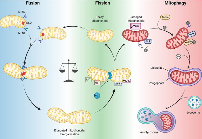
Fusion: This process allows for mitochondria to join together and share contents. This helps to dilute any minor damage by sharing proteins, lipids, and DNA. Key proteins involved in fusion are:
MFN-1 and MFN-2 - these proteins are located on the outside of the mitochondria and act like docking stations to join mitochondria together and allow for the mixing of lipids, proteins and distribution of DNA.⁸ MFN-2 is also used to help tether mitochondria to the endoplasmic reticulum (ER). A decline in MFN-2 has been associated with fragmented mitochondria, poor energy production, and even insulin resistance.⁹
OPA1 - reduced OPA1 and Mitofusion activity is associated with aging, neurodegeneration, and metabolic dysfunction.¹⁰
Fission: Mitochondria can split themselves up, allowing for damaged sections of mitochondria to be removed from the mitochondrial network. Without fission, damaged mitochondria would accumulate, mitophagy couldn't occur, and cellular stress would increase. Key proteins include DRP1 (dynamin-related protein 1), FIS1, MFF (mitochondrial fission factor), and MID49/MID51.¹¹
Mitophagy: This process is similar to autophagy; mitochondria that have been severely damaged and are no longer function are targeted and recycled; their components can go on to be used by the remaining mitochondrial pool. Mitophagy can be triggered by multiple pathways including PINK/PARKIN and BNIP3/NIX.¹² The PINK/Parkin pathway detects dysfunctional mitochondria and labels them for degradation. Defects in the PINK1-Parkin pathway are among the most well-characterised examples of how impaired mitochondrial quality control contributes to neurodegenerative disease including Parkinson's.¹³
Healthy ageing depends on a continuous cycle of mitochondrial maintenance. OPA1 helps mitochondria share resources, DRP1 separates damaged components, PINK1 and Parkin remove dysfunctional mitochondria, and PGC-1α helps build new ones. Together these systems allow cells to adapt to stress and maintain energy production over time. As we age, this quality-control network gradually becomes less efficient, leading to the accumulation of dysfunctional mitochondria that contribute to many of the hallmarks of ageing.
What happens during exercise?
During exercise, working muscles demand far more ATP than they do at rest, and mitochondria respond by ramping up electron transport chain activity. This temporarily increases ROS output, which, as covered above, acts as a signal rather than a threat at these levels. One of the key pathways activated is PGC1-a, often described as the "master regulator" of mitochondrial biogenesis, which switches on the creation of new mitochondria and encourages the existing network to become more efficient.⁵
Exercise also activates AMPK, the cell's energy-sensing pathway, which is triggered as ATP is used up faster than it's replenished.¹⁴ AMPK helps redirect the cell towards energy-producing processes and supports the same quality-control systems discussed earlier, including mitophagy, to clear out mitochondria that aren't keeping up. Over repeated bouts of exercise, this cycle of stress and adaptation is what drives the improvements in endurance, metabolic flexibility, and mitochondrial density associated with regular training.
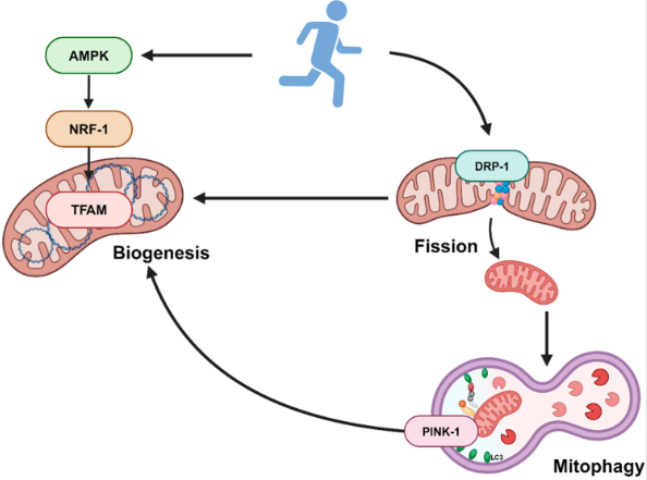
Exercise activates AMPK to drive new mitochondrial biogenesis (via NRF-1 and TFAM), while also triggering fission (DRP-1) and mitophagy (PINK-1) to clear out damaged mitochondria - completing the cycle of repair and renewal.
What happens during fasting?
Fasting places a different kind of stress on mitochondria: rather than a sudden spike in energy demand, it's a drop in incoming fuel. As glucose and insulin levels fall, AMPK is activated in a similar way to exercise, signalling to the cell that energy needs to be conserved and generated more efficiently.⁶ This shift also increases NAD+ availability, which activates sirtuins (particularly SIRT1 and SIRT3), a family of proteins closely tied to mitochondrial function, DNA repair, and longevity pathways.⁷
Fasting is also one of the strongest known triggers of autophagy and mitophagy, allowing the cell to clear out damaged components, including underperforming mitochondria, at a faster rate than usual.¹⁵ Combined with the metabolic switch towards fat oxidation and ketone production, this creates a mitochondrial environment that favours quality over quantity, an important part of why intermittent fasting and calorie restriction are studied in the context of healthy aging.⁶
What's the difference between healthy and unhealthy mitochondrial stress?
The distinction largely comes down to intensity, duration, and recovery. Healthy (hormetic) mitochondrial stress is transient: ROS rises briefly, quality-control pathways like fusion, fission, and mitophagy respond, and the cell returns to a stable, often improved, baseline. This is the pattern seen with exercise, fasting, and cold exposure, where a manageable stressor is followed by adequate recovery.
Unhealthy mitochondrial stress occurs when the stressor is too frequent, too intense, or never resolves, so the cell doesn't get the chance to recover between exposures. In this state, ROS production can outpace the cell's antioxidant defences, quality-control systems can become overwhelmed (damaged mitochondria accumulate faster than mitophagy can clear them), and chronic low-grade oxidative damage to lipids, proteins, and mitochondrial DNA can build up over time.²,³ This is the type of mitochondrial stress that has been linked to accelerated aging and a range of chronic health conditions, rather than the adaptive stress associated with exercise or fasting.
Mitoquinol (MitoQ) and the cellular stress response
Because mitochondria-targeted antioxidants like MitoQ accumulate specifically within the mitochondria rather than acting broadly throughout the body,¹ they're positioned to support the cell's redox balance at the exact site where ROS is produced during stress. This targeted approach is different to how high-dose, non-targeted antioxidants (like large amounts of vitamin C or E) work, which some research suggests may blunt the beneficial signalling role of ROS if taken around exercise, potentially interfering with the very adaptations that hormetic stress is meant to trigger.¹⁶,¹⁷
The proposed role of MitoQ within the cellular stress response isn't to eliminate ROS altogether, but to help keep oxidative stress within a manageable range, supporting mitochondrial quality-control processes like fusion, fission, and mitophagy to function as intended, rather than becoming overwhelmed. This is why ongoing research into Mitoquinol spans both everyday contexts (like exercise recovery and healthy aging) and situations of higher mitochondrial stress,¹⁸,¹⁹ exploring whether supporting mitochondrial redox balance can help the body's own adaptive systems do their job more effectively.
References
Murphy MP. How mitochondria produce reactive oxygen species. Biochemical Journal. 2009;417(1):1-13. DOI: https://doi.org/10.1042/BJ20081386
Balaban RS, Nemoto S, Finkel T. Mitochondria, oxidants, and aging. Cell. 2005;120(4):483-495. DOI: https://doi.org/10.1016/j.cell.2005.02.001
Brand MD. The sites and topology of mitochondrial superoxide production. Experimental Gerontology. 2010;45(7-8):466-472. DOI: https://doi.org/10.1016/j.exger.2010.01.003
Merry TL, Ristow M. Do antioxidant supplements interfere with skeletal muscle adaptation? The Journal of Physiology. 2016;594(18):5135-5147. DOI: https://doi.org/10.1113/JP270654
Handschin C, Spiegelman BM. Peroxisome proliferator-activated receptor gamma coactivator 1 coactivators, energy homeostasis, and metabolism. Endocrine Reviews. 2006;27(7):728-735. DOI: https://doi.org/10.1210/er.2006-0037
Longo VD, Mattson MP. Fasting: molecular mechanisms and clinical applications. Cell Metabolism. 2014;19(2):181-192. DOI: https://doi.org/10.1016/j.cmet.2013.12.008
Verdin E. NAD+ in aging, metabolism, and neurodegeneration. Science. 2015;350(6265):1208-1213. DOI: https://doi.org/10.1126/science.aac4854
Chen H, Chan DC. Mitochondrial dynamics-fusion, fission, movement, and mitophagy-in neurodegenerative diseases. Human Molecular Genetics. 2009;18(R2):R169-R176. DOI: https://doi.org/10.1093/hmg/ddp326
Sebastián D, Hernández-Alvarez MI, Segalés J, et al. Mitofusin 2 (Mfn2) links mitochondrial and endoplasmic reticulum function with insulin signaling and is essential for normal glucose homeostasis. PNAS. 2012;109(14):5523-5528. DOI: https://doi.org/10.1073/pnas.1108220109
MacVicar T, Langer T. OPA1 processing in cell death and disease: the long and short of it. Journal of Cell Science. 2016;129(12):2297-2306. DOI: https://doi.org/10.1242/jcs.159186
Kraus F, Ryan MT. The constriction and scission machineries involved in mitochondrial fission. Journal of Cell Science. 2017;130(18):2953-2960. DOI: https://doi.org/10.1242/jcs.199562
Pickles S, Vigié P, Youle RJ. Mitophagy and quality control mechanisms in mitochondrial maintenance. Current Biology. 2018;28(4):R170-R185. DOI: https://doi.org/10.1016/j.cub.2018.01.004
Pickrell AM, Youle RJ. The roles of PINK1, parkin, and mitochondrial fidelity in Parkinson's disease. Neuron. 2015;85(2):257-273. DOI: https://doi.org/10.1016/j.neuron.2014.12.007
Richter EA, Ruderman NB. AMPK and the biochemistry of exercise: implications for human health and disease. Biochemical Journal. 2009;418(2):261-275. DOI: https://doi.org/10.1042/BJ20082055
Alirezaei M, Kemball CC, Flynn CT, Wood MR, Whitton JL, Kiosses WB. Short-term fasting induces profound neuronal autophagy. Autophagy. 2010;6(6):702-710. DOI: https://doi.org/10.4161/auto.6.6.12376
Ristow M, Zarse K, Oberbach A, et al. Antioxidants prevent health-promoting effects of physical exercise in humans. PNAS. 2009;106(21):8665-8670. DOI: https://doi.org/10.1073/pnas.0903485106
Paulsen G, Cumming KT, Holden G, et al. Vitamin C and E supplementation hampers cellular adaptation to endurance training in humans: a double-blind, randomised, controlled trial. The Journal of Physiology. 2014;592(8):1887-1901. DOI: https://doi.org/10.1113/jphysiol.2013.267419
Rossman MJ, Santos-Parker JR, Steward CAC, et al. Chronic supplementation with a mitochondrial antioxidant (MitoQ) improves vascular function in healthy older adults. Hypertension. 2018;71(6):1056-1063. DOI: https://doi.org/10.1161/HYPERTENSIONAHA.117.10787
Broome SC, Woodhead JST, Merry TL, et al. Mitochondria-targeted antioxidant supplementation improves 8 km time trial performance. Journal of the International Society of Sports Nutrition. 2021;18:58. DOI: https://doi.org/10.1186/s12970-021-00454-0