Mitochondrial Dysfunction
When Cellular Energy, Signalling, and Quality Control Lose Resilience
An Overview of Mitochondrial Dysfunction
Mitochondrial dysfunction is often described as an energy problem, but it is better understood as a loss of mitochondrial performance or adaptability. Mitochondria must match ATP production to changing demand while coordinating substrate metabolism, redox and calcium signalling, innate immunity, cell-fate decisions, and the maintenance of mitochondrial proteins and DNA.¹⁻⁴ Dysfunction becomes clinically relevant when these systems can no longer sustain tissue needs or recover effectively from stress.
This article focuses on mitochondrial dysfunction encountered in the context of aging, chronic disease, inflammation, metabolic stress, medication exposure, or complex presentations such as fatigue and reduced exercise tolerance. This differs from primary mitochondrial disease, an inherited group of disorders caused by pathogenic variants in mitochondrial or nuclear DNA that directly impair mitochondrial structure or function and usually require specialist genetic assessment.⁵,⁶
| Concept | Summary |
|---|---|
| The central problem | Mitochondria must sustain cellular function while demand, nutrient availability, and stress continually change. |
| What dysfunction means | Loss of performance or adaptability across energy production, signalling, dynamics, genome integrity, or quality control. |
| Where it matters | Most evident in tissues with high or fluctuating energy demands, including brain, heart, vascular tissue, and skeletal muscle. |
| Associated conditions | Aging, neurodegenerative disease, insulin resistance and type 2 diabetes, cardiovascular dysfunction, frailty, and sarcopenia. |
| How it is measured | No single test is sufficient; strong studies combine molecular or mitochondrial measures with functional outcomes. |
| Most established support | Exercise has the strongest human evidence for coordinated mitochondrial remodelling and adaptation. |
| Core caution | A mitochondrial biomarker can show target engagement without demonstrating symptom, disease, healthspan, or lifespan benefit. |
Why Mitochondrial Dysfunction Matters in Clinical Practice
Clinicians rarely see “mitochondrial dysfunction” as an isolated diagnosis. More often, impaired mitochondrial biology may contribute to the symptom burden, progression, or reduced resilience associated with an established condition. A patient with type 2 diabetes may show impaired fuel switching and altered substrate oxidation; an older adult with sarcopenia may have reduced respiratory reserve and mitochondrial turnover; and a patient with a neurodegenerative disorder may show disruption of mitochondrial transport, calcium control, or mitophagy.⁷⁻¹⁰
The same framework may be relevant in complex presentations involving persistent fatigue, exercise intolerance, slow recovery, or cognitive symptoms. Fatigue has many possible causes, and evidence linking specific mitochondrial abnormalities to fatigue remains heterogeneous. It is therefore appropriate to consider mitochondrial function as one possible contributor only after common medical, sleep-related, nutritional, psychological, medication-related, endocrine, infectious, and cardiopulmonary explanations have been assessed.¹¹
The practical question is not whether a patient can be labelled with mitochondrial dysfunction. It is whether impaired cellular energy handling, redox control, or mitochondrial quality may help explain the pattern being observed and whether this changes assessment, management of the underlying condition, or interpretation of treatment response.
What Changes When Mitochondrial Function Declines?
Bioenergetic Reserve and Metabolic Flexibility
Mitochondrial impairment may involve reduced electron-transport-chain activity, weaker coupling between oxygen consumption and ATP synthesis, altered substrate oxidation, or loss of spare respiratory capacity. Resting ATP can remain relatively stable because cells compensate through glycolysis or altered fuel use. The limitation may become apparent only when demand rises during exercise, illness, inflammation, or recovery.¹,⁷
Redox and Calcium Regulation
Reactive oxygen species are normal signalling molecules, not simply waste products. Problems arise when their amount, location, duration, or clearance is no longer controlled. Persistent oxidant exposure can damage proteins, lipids, and DNA, while excessive mitochondrial calcium uptake can disrupt membrane potential and promote cell injury.²,³ Oxidative stress may accompany mitochondrial dysfunction, but it is not equivalent to it and may arise from non-mitochondrial sources.
Quality Control, Dynamics and Genome Maintenance
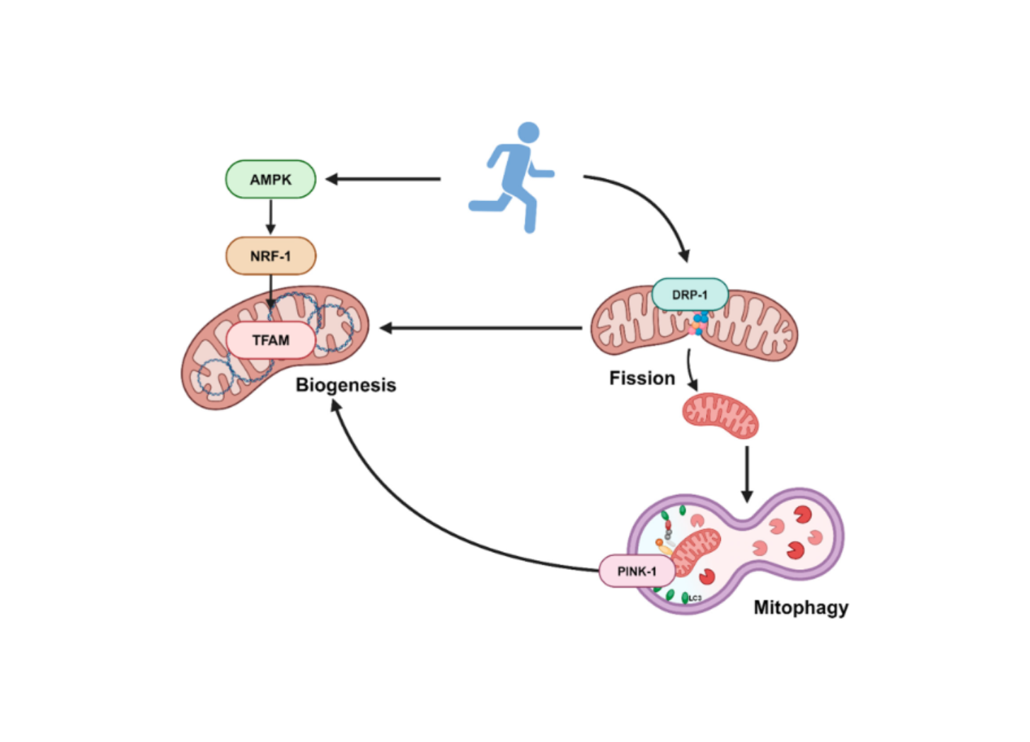
Mitochondria are maintained through proteases and chaperones, fusion and fission, mitophagy, and biogenesis. Fusion supports exchange within the network; fission helps distribution and separation of damaged regions; mitophagy removes mitochondria that cannot be recovered; and biogenesis replenishes capacity.⁴,¹² Dysfunction develops when repair, removal, and replacement no longer keep pace with damage. Mutations or deletions in mitochondrial DNA and disruption of mitochondrial protein import or folding may further impair respiratory-chain assembly and cellular stress responses.⁵,¹³
Why Mitochondrial Dysfunction Increases With Age
Aging does not simply reduce mitochondrial number. It changes the coordination of maintenance and adaptation. Mitophagy and proteostasis may become less efficient, mitochondrial DNA mutations can expand within tissues, and nutrient- and energy-sensing pathways change. Biogenesis may still occur, but increasing quantity does not restore function if damaged mitochondria are not removed or replacement components are poorly assembled.¹²⁻¹⁴
This can create a self-reinforcing cycle. Damaged mitochondria may reduce respiratory reserve, destabilise redox and calcium regulation, and release mitochondrial DNA or other components that activate innate immune pathways. Inflammation then places further pressure on mitochondrial metabolism and turnover. Mitochondrial dysfunction also interacts with cellular senescence, impaired autophagy, loss of proteostasis, deregulated nutrient sensing, and other hallmarks of aging.¹⁴⁻¹⁶
Clinical Contexts in Which Mitochondrial Dysfunction May Be Relevant
Cardiometabolic Disease
Mitochondria influence glucose and fatty-acid oxidation, lipid handling, and metabolic flexibility. Altered respiratory function, substrate use, redox balance, and quality control are reported in insulin resistance, type 2 diabetes, metabolic syndrome, fatty liver disease, and cardiovascular disease.⁷,⁸ These findings interact with nutrient excess, inactivity, inflammation, vascular factors, and disease stage; they should not be interpreted as one universal mitochondrial defect.
Neurodegenerative and Musculoskeletal Conditions
Neurons rely on continuous energy supply, mitochondrial transport, calcium regulation, and mitophagy. Disturbances in these processes are widely studied in Alzheimer and Parkinson diseases.⁹ Skeletal muscle requires mitochondrial reserve for activity and recovery, and age-related changes in respiration and turnover may interact with inactivity, inflammation, vascular impairment, undernutrition, and anabolic resistance in sarcopenia and frailty.¹⁰
Fatigue, Exercise Intolerance and Reduced Recovery
Mitochondrial dysfunction is biologically plausible in some fatigue states because impaired ATP production, fatty-acid metabolism, redox regulation, or inflammatory signalling could reduce endurance or prolong recovery. However, studies use varied populations and biomarkers, and fatigue remains nonspecific.¹¹ A mitochondrial perspective is most useful when symptoms are persistent, disproportionate to activity, associated with reduced functional capacity, or occur alongside a condition already linked to altered mitochondrial biology. It should complement, not replace, standard differential diagnosis.
When Should Clinicians Consider Mitochondrial Dysfunction?
Consider mitochondrial involvement when a patient has an established condition associated with altered mitochondrial biology and also shows reduced exercise tolerance, unexplained decline in physical capacity, poor recovery, metabolic inflexibility, or persistent fatigue. It may also be relevant when multiple systems are affected, symptoms worsen under physiological stress, or conventional assessment does not fully explain the functional impairment. These features are prompts for broader evaluation, not diagnostic criteria.
Primary mitochondrial disease requires a different level of concern. Early onset, maternal or suggestive family history, progressive multisystem involvement, neurological regression, seizures, hearing or visual loss, cardiomyopathy, marked muscle weakness, recurrent metabolic decompensation, or other characteristic phenotypes should prompt referral to a mitochondrial, metabolic, neurology, or genetics service. Genetic testing is central to molecular confirmation, supported by phenotype-directed biochemical or tissue investigations.⁵,⁶
How Can Mitochondrial Function Be Explored?
There is no single validated test for mitochondrial dysfunction associated with aging or common chronic disease. Clinicians should begin with the underlying condition, medication and exposure history, nutritional status, sleep, activity, functional capacity, and routine investigations relevant to the presentation. Selected metabolic, oxidative-damage, and antioxidant-status measures may add context, but most do not measure mitochondrial function directly. Specialist biomarkers and genetic testing are mainly relevant when primary mitochondrial disease is suspected.⁶,¹⁷
Common Measures Used in Clinical and Research Settings
| Measure | What it may show | When it may help | Setting |
|---|---|---|---|
| Routine pathology and metabolic assessment | Alternative or contributing causes of fatigue, poor recovery, or metabolic dysfunction | Initial assessment guided by the patient’s symptoms and known conditions | Clinical |
| Lactate, pyruvate and metabolic profiles | Patterns suggesting altered oxidative or intermediary metabolism; findings are nonspecific | Phenotype suggesting primary mitochondrial or metabolic disease | Specialist clinical |
| F2-isoprostanes and 8-OHdG | Lipid and DNA oxidation, respectively; neither identifies mitochondria as the source | Characterising oxidative burden in a defined clinical or research question | Clinical / research |
| Glutathione and antioxidant-enzyme measures | Context on redox and antioxidant status; results are sample- and assay-dependent | Redox assessment or intervention monitoring | Clinical / research |
| GDF-15 and FGF-21 | Supportive stress biomarkers studied mainly in primary mitochondrial disease | Specialist investigation of a compatible phenotype | Specialist clinical |
| Genetic testing | Pathogenic mtDNA or nuclear DNA variants | Suspected inherited mitochondrial disease | Specialist genetics |
Supporting Mitochondrial Resilience
Exercise provides the strongest human evidence for coordinated mitochondrial adaptation. Aerobic, interval, and resistance exercise can influence mitochondrial biogenesis, respiratory capacity, substrate use, redox regulation, and turnover, while also improving clinically meaningful outcomes such as aerobic capacity, muscle performance, and metabolic health.¹⁰,²¹ The programme should be matched to diagnosis, baseline capacity, symptoms, and recovery, especially when exertion worsens the presentation.
Adequate sleep, management of cardiometabolic risk, smoking avoidance, and sufficient energy, protein, and micronutrient intake support general physiological resilience. These measures should not be presented as specific treatments for mitochondrial dysfunction. Nutrients or supplements should be evaluated by formulation, dose, population, duration, mitochondrial endpoint, functional outcome, safety, and potential interactions.
How Does MitoQ Mitoquinol Support Mitochondrial Function?
MitoQ Mitoquinol is a clinically studied, mitochondria-targeted form of coenzyme Q10. Its triphenylphosphonium component drives accumulation within mitochondria, positioning the antioxidant ubiquinol component near the inner mitochondrial membrane and respiratory chain. Once oxidised, Mitoquinol can be recycled to its active antioxidant form, supporting continued activity within the organelle.²²
This targeted mechanism enables Mitoquinol to support several processes affected by mitochondrial dysfunction. In a double-blind, randomised, placebo-controlled study of 24 healthy participants, 20 mg/day for 21 days attenuated exercise-induced mitochondrial and nuclear DNA damage in lymphocytes and skeletal muscle.²³ Human skeletal-muscle research has also reported reduced mitochondrial hydrogen-peroxide emission and increased catalase protein expression following supplementation.²⁴
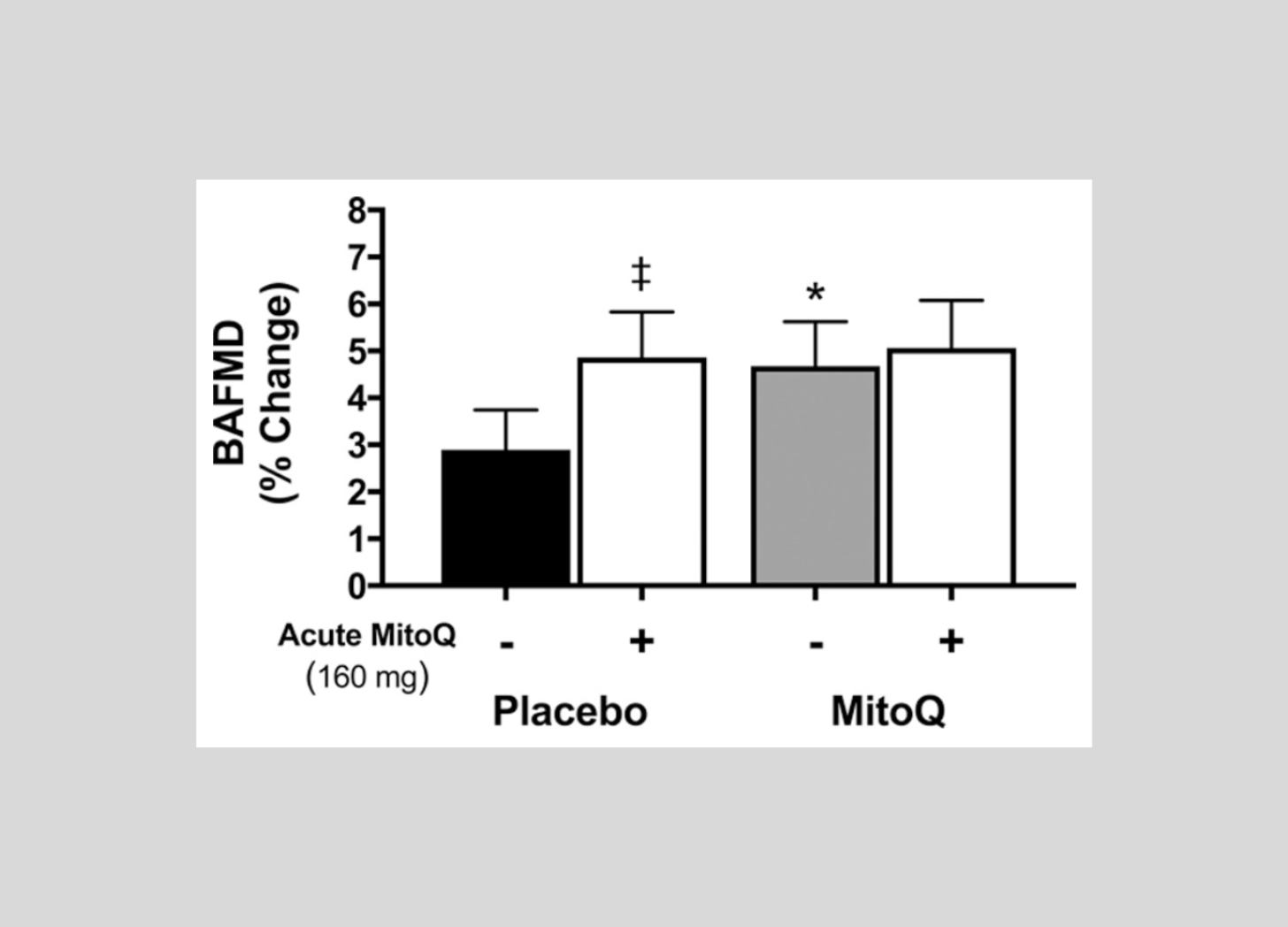
These cellular effects are supported by clinically relevant outcomes. Six weeks of Mitoquinol supplementation improved endothelial function in older adults with impaired vascular function, while exercise studies have reported modulation of PGC-1α signalling and selected performance outcomes.²⁵,²⁶ A 2024 meta-analysis of eight studies involving 188 participants found that Mitoquinol significantly reduced exercise-induced oxidative damage.²⁷ Together, the evidence positions Mitoquinol as a targeted strategy for supporting mitochondrial redox balance, protecting mitochondrial and nuclear DNA, strengthening endogenous antioxidant defences, and maintaining redox-sensitive physiological function.
Key Takeaways
Mitochondrial dysfunction is a loss of mitochondrial performance or adaptability, not a single diagnosis.
For most clinicians, it is relevant as a potential contributor to fatigue, poor exercise tolerance, reduced recovery, or progression within an established condition.
Primary mitochondrial disease is an inherited disorder requiring specialist clinical and genetic assessment; it is not the main focus of this article.
No single test confirms mitochondrial dysfunction associated with aging or common chronic disease.
Oxidative-stress and antioxidant markers provide context but do not identify mitochondria as the source.
Clinical interpretation should integrate the underlying condition, differential diagnosis, functional capacity, relevant biomarkers, and response to evidence-based care.
References
Spinelli JB, Haigis MC. The multifaceted contributions of mitochondria to cellular metabolism. Nat Cell Biol. 2018;20(7):745-754. doi:10.1038/s41556-018-0124-1.
Sena LA, Chandel NS. Physiological roles of mitochondrial reactive oxygen species. Mol Cell. 2012;48(2):158-167. doi:10.1016/j.molcel.2012.09.025.
Rizzuto R, De Stefani D, Raffaello A, Mammucari C. Mitochondria as sensors and regulators of calcium signalling. Nat Rev Mol Cell Biol. 2012;13(9):566-578. doi:10.1038/nrm3412.
Pickles S, Vigie P, Youle RJ. Mitophagy and quality-control mechanisms in mitochondrial maintenance. Curr Biol. 2018;28(4):R170-R185. doi:10.1016/j.cub.2018.01.004.
Niyazov DM, Kahler SG, Frye RE. Primary mitochondrial disease and secondary mitochondrial dysfunction: importance of distinction for diagnosis and treatment. Mol Syndromol. 2016;7(3):122-137. doi:10.1159/000446586.
Kose M, McCormick E, Peterson JT, et al. Advances in diagnosis of mitochondrial dysfunction and primary mitochondrial disease. Dis Model Mech. 2026;19:dmm052804. doi:10.1242/dmm.052804.
Montgomery MK, Turner N. Mitochondrial dysfunction and insulin resistance: an update. Endocr Connect. 2015;4(1):R1-R15. doi:10.1530/EC-14-0092.
Pinti MV, Fink GK, Hathaway QA, et al. Mitochondrial dysfunction in type 2 diabetes mellitus: an organ-based analysis. Am J Physiol Endocrinol Metab. 2019;316(2):E268-E285. doi:10.1152/ajpendo.00314.2018.
Swerdlow RH. Mitochondria and mitochondrial cascades in Alzheimer disease. J Alzheimers Dis. 2018;62(3):1403-1416. doi:10.3233/JAD-170585.
Coen PM, Musci RV, Hinkley JM, Miller BF. Mitochondria as a target for mitigating sarcopenia. Front Physiol. 2019;9:1883. doi:10.3389/fphys.2018.01883.
Filler K, Lyon D, Bennett J, et al. Association of mitochondrial dysfunction and fatigue: a review of the literature. BBA Clin. 2014;1:12-23. doi:10.1016/j.bbacli.2014.04.001.
Palikaras K, Lionaki E, Tavernarakis N. Mechanisms of mitophagy in cellular homeostasis, physiology and pathology. Nat Cell Biol. 2018;20(9):1013-1022. doi:10.1038/s41556-018-0176-2.
Quiros PM, Langer T, Lopez-Otin C. New roles for mitochondrial proteases in health, ageing and disease. Nat Rev Mol Cell Biol. 2015;16(6):345-359. doi:10.1038/nrm3984.
Sun N, Youle RJ, Finkel T. The mitochondrial basis of aging. Mol Cell. 2016;61(5):654-666. doi:10.1016/j.molcel.2016.01.028.
Riley JS, Tait SWG. Mitochondrial DNA in inflammation and immunity. EMBO Rep. 2020;21(4):e49799. doi:10.15252/embr.201949799.
Lopez-Otin C, Blasco MA, Partridge L, Serrano M, Kroemer G. Hallmarks of aging: an expanding universe. Cell. 2023;186(2):243-278. doi:10.1016/j.cell.2022.11.001.
Shayota BJ. Biomarkers of mitochondrial disorders. Neurotherapeutics. 2024;21(1):e00325. doi:10.1016/j.neurot.2024.e00325.
Milatovic D, Montine TJ, Aschner M. Measurement of isoprostanes as markers of oxidative stress. Methods Mol Biol. 2011;758:195-204. doi:10.1007/978-1-61779-170-3_13.
Graille M, Wild P, Sauvain JJ, et al. Urinary 8-OHdG as a biomarker for oxidative stress: a systematic literature review and meta-analysis. Int J Mol Sci. 2020;21(11):3743. doi:10.3390/ijms21113743.
Silvestrini A, Meucci E, Ricerca BM, Mancini A. Total antioxidant capacity: biochemical aspects and clinical significance. Int J Mol Sci. 2023;24(13):10978. doi:10.3390/ijms241310978.
Granata C, Jamnick NA, Bishop DJ. Principles of exercise prescription and mitochondrial biogenesis. Sports Med. 2018;48(7):1541-1559. doi:10.1007/s40279-018-0894-4.
Murphy MP, Smith RAJ. Targeting antioxidants to mitochondria by conjugation to lipophilic cations. Annu Rev Pharmacol Toxicol. 2007;47:629-656. doi:10.1146/annurev.pharmtox.47.120505.105110.
Williamson J, Hughes CM, Cobley JN, Davison GW. The mitochondria-targeted antioxidant MitoQ attenuates exercise-induced mitochondrial DNA damage. Redox Biol. 2020;36:101673. doi:10.1016/j.redox.2020.101673.
Pham T, MacRae CL, Broome SC, et al. MitoQ and CoQ10 supplementation mildly suppress skeletal muscle mitochondrial hydrogen peroxide levels without impacting mitochondrial function in middle-aged men. Eur J Appl Physiol. 2020;120:1657-1669. doi:10.1007/s00421-020-04396-4.
Rossman MJ, Santos-Parker JR, Steward CAC, et al. Chronic supplementation with a mitochondrial antioxidant (MitoQ) improves vascular function in healthy older adults. Hypertension. 2018;71(6):1056-1063. doi:10.1161/HYPERTENSIONAHA.117.10787.
Broome SC, Pham T, Braakhuis AJ, et al. MitoQ supplementation augments acute exercise-induced increases in muscle PGC-1α mRNA and improves training-induced increases in peak power independent of mitochondrial content and function in untrained middle-aged men. Redox Biol. 2022;53:102341. doi:10.1016/j.redox.2022.102341.
Gonzalo-Skok O, et al. Effects of Mitoquinone (MitoQ) supplementation on aerobic exercise performance and oxidative damage: a systematic review and meta-analysis. Sports Med Open. 2024;10:80. doi:10.1186/s40798-024-00741-5.
Latest Resources
Prescriptions







MitoQ (Mitoquinol Mesylate)
1000+ peer-reviewed articles
30+ Clinical Trials